
Chapter: Female Pelvis
Introduction:
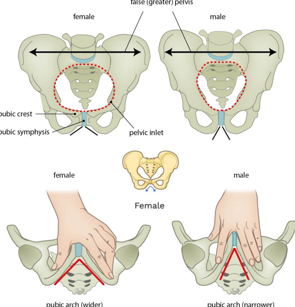
The pelvis is a bony structure of the skeletal system that plays a crucial role in supporting body movement and posture. In females, it is specially adapted for childbearing. Compared to males, the female pelvis is wider, more circular, and designed to support pregnancy and childbirth.
Structure of the Pelvis:
The pelvis is made up of four bones:
- Two Innominate (Hip) Bones
- One Sacrum
- One Coccyx
1. Innominate Bones (Hip Bones):
Each innominate bone is formed by the fusion of three bones:
a. Ilium:
- The upper and outer portion of the hip bone.
- The Iliac Crest is the prominent ridge felt when placing hands on the hips.
- Anterior superior iliac spine and anterior inferior iliac spine are important anatomical landmarks.
b. Ischium:
- The lower, thick, and strong part of the hip bone.
- Contains the Ischial tuberosity, which supports the body while sitting.
- Ischial spines are projections important during childbirth to assess fetal head position.
c. Pubis:
- The front part of the hip bone.
- Two pubic bones join at the pubic symphysis.
- Consists of superior and inferior pubic ramus.
2. Sacrum:
- A triangular-shaped bone formed by the fusion of five sacral vertebrae.
- The sacral promontory is the uppermost part.
- Located at the back of the pelvis, connecting with the iliac bones at the sacroiliac joints.
3. Coccyx:
- Known as the tailbone, it is a small, curved bone at the base of the spine.
- Formed by the fusion of four coccygeal vertebrae.
Pelvic Joints:
The bones of the pelvis are joined together at four joints:
- Two Sacroiliac joints
- One Sacrococcygeal joint
- One Symphysis pubis
Divisions of the Pelvis:
1. False Pelvis (Greater Pelvis):
- Formed by the upper part of the hip bones.
- It does not participate in childbirth.
- It provides support to the uterus during pregnancy.
2. True Pelvis (Lesser Pelvis):
- This is the functional part involved in childbirth.
- Divided into three regions:
- Pelvic Inlet (Brim)
- Pelvic Cavity (Mid-pelvis)
- Pelvic Outlet
Pelvic Ligaments:
- Sacroiliac ligament
- Sacrococcygeal ligament
- Pubic ligament
- Sacrotuberous ligament
- Sacrospinous ligament
Pelvic Landmarks:
- Sacral Promontory
- Ala of Sacrum
- Sacroiliac Joint
- Iliopectineal Line
- Iliopubic Eminence
- Pectineal Line
- Pubic Tubercle
- Pubic Crest
- Symphysis Pubis
Diameters of Female Pelvis:
| Region | Anteroposterior Diameter | Transverse Diameter | Oblique Diameter |
|---|---|---|---|
| Inlet | 11 cm | 13 cm | 12 cm |
| Cavity | 12 cm | 12 cm | 12 cm |
| Outlet | 13 cm | 11 cm | ~12 cm |
Pelvic Inlet Diameters:
- Anteroposterior: From sacral promontory to upper border of symphysis pubis = 11 cm
- Transverse: Between iliopectineal lines = 13 cm
- Oblique: From sacroiliac joint to iliopectineal eminence = 12 cm
Pelvic Cavity Diameters:
- Considered almost circular.
- All diameters (anteroposterior, transverse, oblique) ≈ 12 cm
Pelvic Outlet Diameters:
- Anteroposterior: From tip of coccyx to lower border of symphysis pubis = 13 cm
- Transverse: Between ischial spines = 11 cm
- Oblique: Not precisely measured, but approx. 12 cm
Conclusion:
The female pelvis is specially designed to support pregnancy and childbirth. Its anatomical features, diameters, and flexibility are crucial for safe delivery. A proper understanding of the pelvic structure is essential for nursing and medical professionals.