
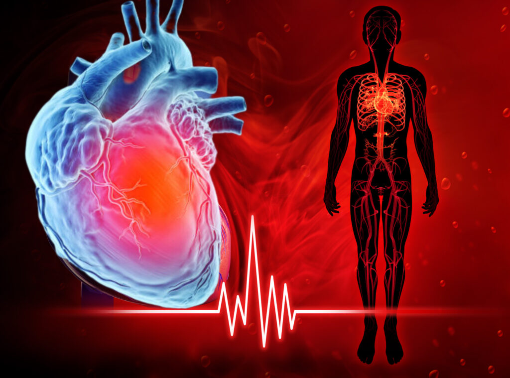
1. Introduction to the Heart
- Function: The heart is a muscular organ that pumps blood throughout the body via the circulatory system.
- Location: Between the lungs in the thoracic cavity, slightly tilted to the left.
- Size: Approximately the size of a clenched fist.
- Layers:
- Pericardium (Outer protective sac)
- Myocardium (Muscular middle layer)
- Endocardium (Inner lining)
2. Chambers of the Heart
The heart has four chambers:
| Chamber | Function |
|---|---|
| Right Atrium | Receives deoxygenated blood from the body via the superior & inferior vena cava. |
| Right Ventricle | Pumps deoxygenated blood to the lungs via the pulmonary artery. |
| Left Atrium | Receives oxygenated blood from the lungs via the pulmonary veins. |
| Left Ventricle | Pumps oxygenated blood to the entire body via the aorta (thickest wall for high pressure). |
Note:
- The left ventricle has the thickest myocardium because it pumps blood to the entire body.
- The right ventricle has thinner walls since it only pumps blood to the lungs.
3. Major Blood Vessels
| Vessel | Function |
|---|---|
| Superior Vena Cava | Brings deoxygenated blood from the upper body to the right atrium. |
| Inferior Vena Cava | Brings deoxygenated blood from the lower body to the right atrium. |
| Pulmonary Artery | Carries deoxygenated blood from the right ventricle to the lungs. (Only artery with deoxygenated blood!) |
| Pulmonary Veins | Bring oxygenated blood from the lungs to the left atrium. (Only veins with oxygenated blood!) |
| Aorta | Distributes oxygenated blood from the left ventricle to the entire body. |
4. Heart Valves (Ensure One-Way Blood Flow)
The heart has four valves to prevent backflow:
| Valve | Location | Function |
|---|---|---|
| Tricuspid Valve | Between right atrium & right ventricle | Prevents backflow into the right atrium. |
| Pulmonary Valve | Between right ventricle & pulmonary artery | Prevents backflow into the right ventricle. |
| Mitral (Bicuspid) Valve | Between left atrium & left ventricle | Prevents backflow into the left atrium. |
| Aortic Valve | Between left ventricle & aorta | Prevents backflow into the left ventricle. |
Mnemonic for Valve Order:
“Try Pulling My Aorta” → Tricuspid, Pulmonary, Mitral, Aortic
5. Blood Flow Through the Heart (Step-by-Step)
- Deoxygenated Blood enters the right atrium via the vena cava.
- Passes through the tricuspid valve into the right ventricle.
- The right ventricle pumps blood through the pulmonary valve into the pulmonary artery (to lungs).
- Oxygenation occurs in the lungs.
- Oxygenated Blood returns via pulmonary veins to the left atrium.
- Passes through the mitral valve into the left ventricle.
- The left ventricle pumps blood through the aortic valve into the aorta (to the body).
Visualization:
Body → Vena Cava → Right Atrium → Tricuspid Valve → Right Ventricle → Pulmonary Valve → Lungs → Pulmonary Veins → Left Atrium → Mitral Valve → Left Ventricle → Aortic Valve → Aorta → Body
6. Key Concepts for Exams
✔ Double Circulation: Heart pumps blood to lungs (pulmonary) and body (systemic) separately.
✔ Pulmonary Artery = Only artery with deoxygenated blood.
✔ Pulmonary Veins = Only veins with oxygenated blood.
✔ Left Ventricle = Thickest wall (high-pressure pump).
✔ Valves prevent backflow (lub-dub sound = valves closing).
7. Common Disorders
- Hypertension (High BP): Excess pressure on artery walls.
- Atherosclerosis: Artery blockage due to plaque.
- Heart Attack: Blockage in coronary arteries.
- Valvular Stenosis: Narrowing of heart valves.
Study Tips:
- Draw a labeled diagram of the heart.
- Trace blood flow step-by-step.
- Use flashcards for valves and vessels.